Filariasis is caused by nematodes (roundworms) that inhabit the lymphatics and subcutaneous tissues.
Eight main species infect humans.
Three of these are responsible for most of the morbidity due to filariasis
1- Wuchereria bancrofti
2- Brugia malayi
cause lymphatic filariasis,
3- Onchocerca volvulus
causes onchocerciasis (river blindness).
The other five species are :
- Loa loa,
- Mansonella perstans,
- Mansonella streptocerca.
- Mansonella ozzardi,
- Brugia timori. (The last species also causes lymphatic filariasis.)
Geographic Distribution
- Among the agents of lymphatic filariasis,
- Wuchereria bancrofti is encountered in tropical areas worldwide;
- Brugia malayi is limited to Asia;
- Onchocerca volvulus, the agent of river blindness, occurs mainly in Africa, with additional foci in Latin America and the Middle East.
- Among the other species, Loa loa and
- Mansonella streptocercaare found in Africa;
- Mansonella perstans occurs in both Africa and South America;
- and Mansonella ozzardi occurs only in the American continent.
Morphology
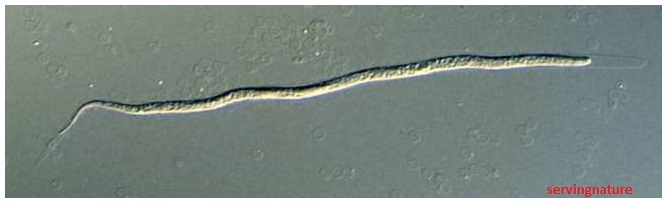
Adult of Wuchereria bancrofti
Adult is milk-white,
Long and slender,
Silk thread-like,
40-100 mm × 0.1-0.2 mm.
live in the lymphatic system.

The female worms measure 80 to 100 mm in length and 0.24 to 0.30 mm in diameter,
while the males measure about 40 mm by 0.1 mm.


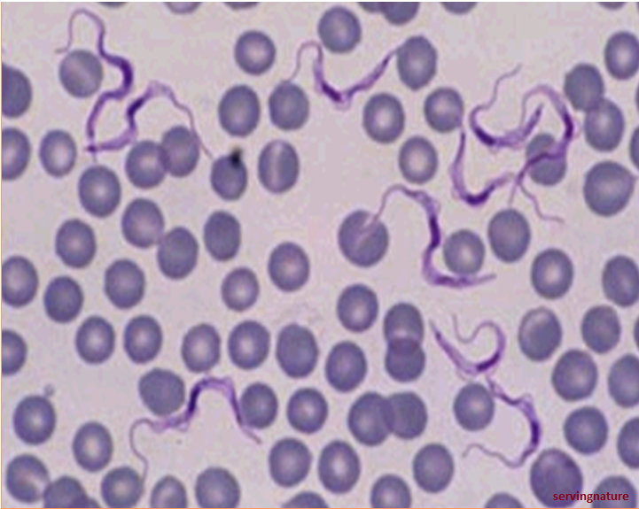
Microfilariae

- The microfilariae of W. bancrofti are sheathed
- Measure 240-230 µm and 275-320 µm in 2% formalin. ( in stained blood smears)
- They have a gently curved body, and a tail that is tapered to a point.
- The nuclear column (the cells that constitute the body of the microfilaria) is loosely packed;
- The cells can be visualized individually and do not extend to the tip of the tail.
- Microfilariae circulate in the blood.

Microfilariae of Brugia malayi


- Microfilariae of Brugia malayi are sheathed
- Measure 175-230 µm or 240-300 µm.
- The tail is tapered, with a significant gap between the terminal and subterminal nuclei.
- Microfilaria circulate in the blood.


Microfilariae of Onchocerca volvulus
- Microfilariae of Onchocerca volvulus are unsheathed
- measure 300-315 µm in length.
- The tail tapers and is often sharply bent.
- The nuclei do not extend to the tip of the tail.
- Microfilariae typically reside in skin but may be found in blood or urine during heavy infections, or invade the eye and cause a condition known as river blindness.

A: Microfilariae of O. volvulus from a skin nodule of a patient
from Zambia, stained with hematoxylin and eosin (H&E).
B: Microfilariae of O. volvulus within the uterus of an adult female.
The specimen was taken from the same patient as in Figure A.

Cross-section of an adult female O. volvulus, stained with H&E.
Note the presence of many microfilariae within the uterus.
Microfilariae of Loa loa
- Microfilariae of Loa loa are sheathed
- Measure 230-250 µm long and 270-300 µm in 2% formalin. in stained blood smears
- The tail is tapered and nuclei extend to the tip of the tail.
- Microfilariae circulate in the blood.


A: Microfilariae of L. loa
Image taken at 100× magnification.
B: Higher magnification of the microfilariae in Figure A,
taken at 500 × oil magnification.
C, D: Adults of L. loa removed from the eye of a patient.
Images courtesy of the Georgia State Public Health Laboratory.
Blackfly, Buffalo gnat, Turkey gnat


Culicoides


Among them are:
- Culex (C. annulirostris, C. bitaeniorhynchus, C. quinquefasciatus, and C. pipiens)
- Anopheles (A. arabinensis, A. bancroftii, A. farauti, A. funestus, A. gambiae, A. koliensis, A. melas, A. merus, A. punctulatus and A. wellcomei)
- Aedes (A. aegypti, A. aquasalis, A. bellator, A. cooki, A. darlingi, A. kochi, A. polynesiensis, A. pseudoscutellaris, A. rotumae, A. scapularis, and A. vigilax)
- Mansonia (M. pseudotitillans, M. uniformis); Coquillettidia (C. juxtamansonia).
- Adults inhabit in lymphatic produce sheathed microfilariae
- The microfilariae migrate into lymph and blood channels moving actively through lymph and blood .
- A mosquito ingests the microfilariae during a blood meal .
- After ingestion, the microfilariae lose their sheaths and some of them work their way through the wall of the proventriculus and cardiac portion of the mosquitoes mid-gut and reach the thoracic muscles .
- There the microfilariae develop into first-stage larvae and subsequently into third-stage infective larvae.
- During a blood meal, an infected mosquito introduces third-stage filarial larvae onto the skin of the human host, where they penetrate into the bite wound .They develop into adults that commonly reside in the lymphatics .
- The third-stage infective larvae migrate through the hemocoele to the mosquitoes proboscis and can infect another human when the mosquito takes a blood meal .
- Adults produce microfilariae measuring 244 to 296 µm by 7.5 to 10 µm, which are sheathed and have nocturnal periodicity, except the South Pacific microfilariae which have the absence of marked periodicity.
- The microfilariae migrate into lymph and blood channels moving actively through lymph and blood .
- The adult worms resemble those of Wuchereria bancrofti but are smaller.
- Female worms measure 43 to 55 mm in length by 130 to 170 µm in width.
- Males measure 13 to 23 mm in length by 70 to 80 µm in width.


Life Cycle of Brugia malayi
The typical vector for Brugia malayi filariasis are mosquito species from the genera Mansonia and Aedes.


Onchocerca Volvulus

Some points to explain
Nocturnal periodicity of microfilariae
(1) Definition
There is a marked periodicity of microfilariae in the peripheral blood, that is, they can be demonstrated at night and disappear during the day.
The microfilariae are concentrated in the small blood vessels of the lungs during the day and are liberated into the peripheral circulation at night. This phenomenon is called nocturnal periodicity of microfilarae.
(2) The time
when the microfilarae reach
the maximal number in the peripheral blood:
W. bancrofti:
between 10 P.M. and 2 A.M.
B. malayi:
between 8 P.M. and 4 A.M.
(3) Mechanism (hypotheses)
A. Suppression of the vagus
(nerve activity)
B. Association with oxygen content
of blood vessels in the lungs.
Microfilaria infectivity to mosquitoes
(1) Species of mosquito vectors of filariae in China For W. bancrofti:
Culex pipiens pallens ,
Anopheles sinensis.
For B. malayi:
Anopheles anthropophagus ,
A. sinensis, and Aedes togoi .
(2) Interaction between microfilariae and mosquitoes:
Fewer than 15 microfilariae / 20 mm3 will fail to infect mosquitoes; that more than 100 will kill the mosquito.
(3) Differences in location in human body between the two filariae.
I) B.malayi:
superficial lymphatics an nodes in arms and legs.
II) W.bancrofti: profound lymphatics and nodes in arms, legs and other parts of the body.
Pathogenesis
- Adult worm are found in lymph vessels throughout the body
- But primarily in or around the axillary, epitrochlear, inguinal and pelvic nodes and the lymphatics dital to them, as well as those of testis, epididymis and cord
Clinical Features
- Lymphatic filariasis most often consists of asymptomatic microfilaremia.
- Some patients develop lymphatic dysfunction causing lymphedema and elephantiasis (frequently in the lower extremities) and, with Wuchereria bancrofti, hydrocele and scrotal elephantiasis.
- Episodes of febrile lymphangitis and lymphadenitis may occur.
1) Microfilaremia
(Asymptomatic phase)
2) Inflammatory (Acute) phase:
intense inflammation of lymphatics and
lymphonoids.
3) Chronic obstructive phase:
elephantiasis, chyluria (lymph in the
urine), hydrocele.
4) Suppressive filariasis (Tropical eosinophilia syndrome)
Symptoms
1) Symptoms in acute phase:
lymphatic inflammation
(1) Susceptible age:
young and strong; Location: legs and arms.
(2) Manifestations:
“retro-lymphangitis”.
W. bancrofti sometimes causes spermatitis,
architis (inflammation of testis) and epididymitis .
(3) Mechanism:
The acute phase commences when the female worms reach maturity and begin releasing miceofilariae. This phase is actually an allergic response to the products of dying and degenerating adult worms.
2) Symptoms in chronic obstructive phase
- Repeated attacks of acute lymphatic inflammations result in a chronic lymphedema with much fibrous infiltration, and consequently lymphatic obstruction.
- The lymph return is obstructed and the lymph “piles up”, greatly dilating the affected duct and forcing the lymph into the surrounding tissues.
(1) Hydrocele, caused by obstruction of lymphatics in testis and spermatic cord.
(2) Chyluria (lymph in urine):
A. common in W. bancrofti filariasis.
B. Manifestations: The chyle gives the urine a milky appearance.
C. Mechanism: preaortic lymph nodes and intestinal lymphatic ducts anterior to chylocystis are obstructed.
(3) Elephantiasis:
A. The most commonly afflicted organs are
scrotum and legs. B. Manifestations C. Mechanism: lymphatic obstruction plus inflammatory hyperplasia

Lymph edema


Elephantiasis
Onchocerca volvulus nodules in scalp

Patient with hanging groin caused by underlying obstruction onchocercal lymphadenitis and loss of dermal elasticity.


Onchocerca volvulus nodules.
Potions of contained adult worms protruding

Moderately advanced elephantiasis involving both legs
With thickening and verrucous,changes of the skin

Extreme elephantiasis of all four limbs and scrotum
Filariasis-Epidemiology
Filariasis is one of the most important parasitic diseases in China.
1) It is distributed in 16 provinces, cities and autonomous regions in the center and south of China.
2) Epidemic factors include:
(1) Source of infection: patients, carriers (infected humans).
(2) Vectors (mosquitoes)
(3) Susceptible population
Filariasis-Prevention and treatment
- Mass examination and treatment
- Mosquito control
- Individual protection Strengthen the epidemiological supervision in the controlled areas.