The definition of anatomy
Anatomy is a science dealing with morphology and structure of the body.
Anatomy includes those structures that can be seen grossly (without the aid of magnification) and microscopically (with the aid of magnification).
Typically, when used by itself, the term 'anatomy' tends to mean gross or macroscopic anatomy-that is, the study of structures that can be seen without using a microscopic.
Microscopic anatomy, also called 'histology', is the study of cells and tissues using a microscope.
The significance of studying anatomy
Anatomy forms the basis for the practice of medicine.
Anatomy leads the physician towards an understanding of a patient's disease whether he or she is carrying out a physical examination or using the most advanced imaging techniques.
Anatomy is also important for dentists, physical therapists, and all others involved in any aspect of patient treatment that begins with an analysis of clinical signs.
The ability to interpret a clinical observation correctly is therefore the endpoint of a sound anatomical understanding.
The classification of anatomy
By the different research methods, anatomy can be divided into micro-anatomy and macro-anatomy.
People recognize human body at different levels.
Whole body---organs(systems)---tissues---cells---cellular organs---ultramicrostructures---molecular
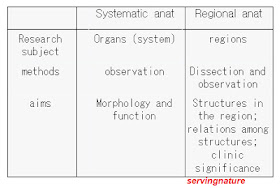
Presently, we study macro-anatomy. According to the different purposes, different research methods, macro-anatomy is divided into systematic anatomy and regional anatomy.
The differences between systematic anatomy and regional anatomy are as follows:
Locomotor system
Protection; support ; movement
Digestive system
To digest foods; to secrete enzymes and hormones( endocrine function ; to absorb the nutrient elements; to eliminate the useless residues
Respiratory system
To supply the blood with oxygen ; to get rid of excess dioxide;
Urinary system
By eliminating the metabolic products to maintain the balance of substances in the body.
Reproductive system
Keeping maintenance of species.
secreting hormone
Circulatory system
Cardiovascular system to transport the substances with oxygen and nutrition.
Lymphatic system
The anatomical position
The body is upright, legs together, and directed forwards
The palms are turned forward, with the thumbs laterally
Anatomical planes
sagittal planes: Refers to any longitudinal
planes that divide the human body into a right and a left part.
Coronal planes: Any vertical planes that divide the body into an anterior and a posterior part
Horizontal (transverse) planes: Any horizontal planes that divide the body into an upper and a lower part.They lie at right angles to both the sagittal and coronal planes.
Terms of direction
Introduction of locomotor system:
The formation of locomotor system, which comprise:
Bones
There are 206 bones in the human body, which can be classified into a number of kinds according to their positions and shapes.
The shape and classifications of bones
1- According to the position,all the bones can be classified into four categories :
skull : 29
bones of trunk: 51
bones of upper limb: 64
bones of lower limb: 62
The structures of bone
1- Periosteum which is a fibrous membrane, containing rich blood vessels and nerves.
Function:
A. play an important role in regeneration of bones, having osteoblast.
B. Provide nutrition for the development, growth and reconstruction of bones
C. Containing receptor (accepting stimulate)
Parts: periosteum (except articular surface) / endosteum
2- Bony substance:
The physical properties of bone
hard: bone removed organic materials; hard but fragile by demonstration;
flexible: bone removed inorganically material specimen, not hard, but flexible
The arrangement of bony substance
A- Compact substance: consist of regular compact bony plate
B- Sponge substance:
outer plate / diploë / inner plate
The arrangement looks like a frame of a house.
Bone marrow
i) Which is located within medullary cavity and ‘network-eye’, divided into red and yellow bone marrow.
ii) 3-5y red bone marrow carrying out the function of blood-forming . For adult most of it become yellow bone marrow, but proximal end of humerus (femur), short bone, flat bone have life-long time red marrow.
outer plate / diploë / inner plate
The structure of bone

Bone is hard and flexible . It depends on its chemical components and arrangements.
Arthrology
Definition: The bones are connected together by means of fibrous, cartilaginous or osseous tissues at different parts of their surface. The connection is called articulation or joint.
1. The classification of articulations
i) Synarthrosis (immovable joints, direct joint)
Definition: two or more separated bones are directly connected by fibrous , cartilaginous or osseous tissues.
Fibrous joints:
a. sutures: skull
b. syndesmoses: ligmentum flava (yellow lig) { tibiofibular joint}
c. Gomphosis
Cartilaginous:
a. synchondrosis: between sternum and 1st costal cartilage
b. symphyses : pubic symphysis
Synosteoses:
Sacrum
ii) Diarthrosis (movable articulations, synovial joints)
Definition: the bones are connected by the joint capsule and ligaments.
A- The essential (basical ) structures of synovial joint
1. The articular surface: which is a part of surface of bone covered by hyaline cartilage.
2. The articular capsule: it looks like a irregular sac, attaches the periphery of the articular surface and adjacent surface. Articular capsule include two parts: outer layer( fibrous layer ) and inner layer( synovial layer---produce synovial fluid)
3. The articular cavity: it is a closed space enclosed by the synovial membrane and the articular cartilage.
B. The accessory structures of the synovial joints
1. The ligment:
intracapsular lig: it is inside the joint , surrounded by synovial membrane\
extracapsular lig: which is outside the capsule
2. The articular disc (or cartilage): it is fibrocartilaginous, and divides the articular cavity partially or completely into 2 parts.
3. The articular lip (labrum): it is a fibrocartilaginous ring, which can deepen the articular surface
C. The movement of joint
1. Flexion and extension: they are performed in the coronal axis. Flexion makes the angle between the adjacent bones decrease; extension increase the angle.
2. Adduction and abduction: which are performed in sagittal axis. Adduction means the movement toward the midline of the body; abduction means the movement apart from the midline.
3. Pronation and supination: in standard anatomical position, the pronation means the palm is turned backward; the supination means the palm is turned forward.
4. Rotation: the movement is performed in the vertical axis. A bone is moving around the vertical axis.
5. Circumduction: while the proximal end of a bone remains relative stable, the distal end moves in a circle.
The movement of joint
Myology
- Skeletal muscle: to move the skeleton
- Cardiac muscle: to form the heart
- Smooth muscle: to constitute viscera
Over 600 muscles in the body
Muscles are grouped by location:
- Muscles of head.
- Muscles of neck.
- Muscles of thorax.
- Muscles of.
- Abdomen.
- Muscles of upper limb.
- Muscles of lower limb.
Naming the skeletal muscles by sereral criteria:
1. location(brachialis);
2. shape (trapezius);
3. direction of the muscle fibers(rectus,oblique);
4. location ofattachments(brachioradialis);
5.number of origins (biceps);
6. action(flexor,extensor)
The structures of muscles
I) Tendon and belly
Belly: the fleshy part of a muscle
Tendon: the bundles consisting of connective tissue blending
with strong collagen
II) The origin and insertion:
Origin: a fixed (less movable) attachment of a muscle
Insertion: a movable attachment of a muscle
Generally speaking, the origin is near the midline; the insertion is far from the midline. However, the origin and insertion may be exchanged each other. For example: pectoralis major m.
III) The relations of muscles to other structures
- Most of muscles are attached to bones,
- some of muscles are attached to skin, e.g; Platysma m. beneath the skin of neck
- some of muscles are attached to organs. e.g; eyeball
IV) The shape of muscles
Because of functional differences, muscles have different shape.
The shape of muscles
IV) The functional classification of muscles
Agonists ( prime movers ): the main muscles which contract to produce desired movement
Antagonist: the muscles which act to oppose the action of agonist
Agonist---contraction; antagonist---relax
Synergist: cooperation in a special action as a supporter
Fixator: fix proximal end of limbs in a special position.
eg. Tightly making a fist
Biceps
Brachialis
Triceps
Fascia and tendinous sheath
Fascia
1) Superficial fascia: lies under skin and covers the entire body containing a lot of fat / increase mobilityof skin; thermal insulation; a store of energy /contain cutaneous nerve, blood vessels and skin muscles
2) Deep fascia: dense and inelastic
membrane of collagenous fibers


No comments:
Post a Comment